
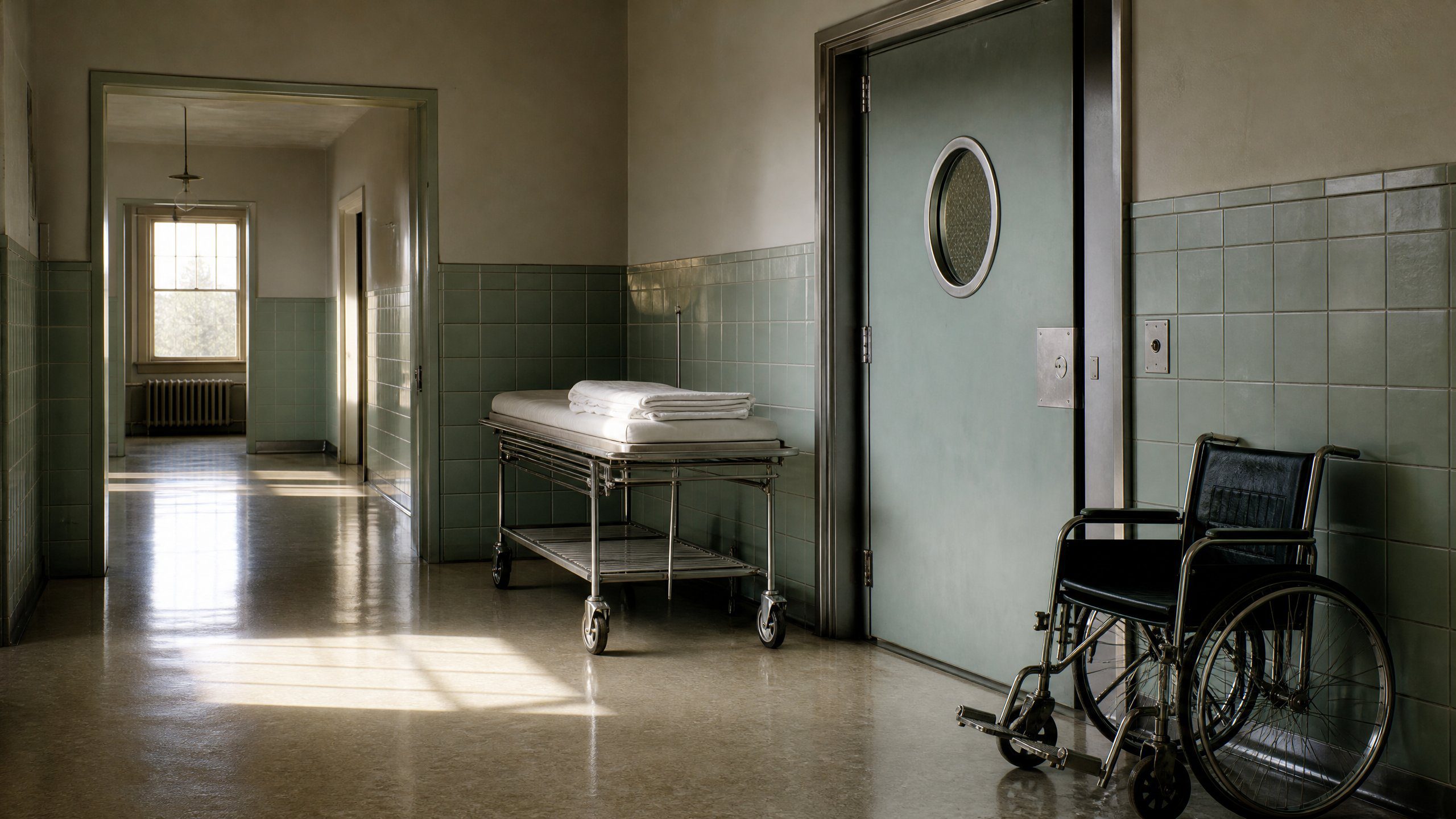
In 1953, a surgeon in Hartford, Connecticut, removed tissue from both sides of a 27-year-old man’s brain to stop his seizures, and the man never formed a lasting new memory again.
The patient was Henry Molaison, and the surgeon was William Beecher Scoville of Hartford Hospital. The operation took out parts of the medial temporal lobes on both sides of the brain, including the hippocampus, and it largely ended the epileptic seizures that had made Molaison’s life unworkable. Afterward he could recall much of his life before 1953, but he could hold on to anything new for only seconds or minutes.
Scientists studied him for more than half a century under the initials H.M., which protected his privacy until his death in 2008. His case showed that the forming of new memories depends on specific structures deep in the temporal lobes, and that the ability to learn a physical skill runs on a separate system that his surgery had left intact.
Henry Molaison’s case
| Surgery | 1953 |
|---|---|
| Surgeon | William Beecher Scoville |
| Age at surgery | 27 |
| Years studied by Corkin | about 46 |
| Died | December 2, 2008, aged 82 |
| Brain slices | 2,401 |
Why did Henry Molaison have brain surgery?
Henry Molaison had brain surgery to stop epileptic seizures that had grown so frequent and violent they made his working life impossible. Molaison grew up in Connecticut, and when he was 9 he was knocked down by a bicycle rider in his neighborhood. He developed seizures afterward, and over the following years they grew more frequent and more violent. He finished high school at 21 and found work as a motor winder on an assembly line, but his blackouts and convulsions eventually made the job impossible.
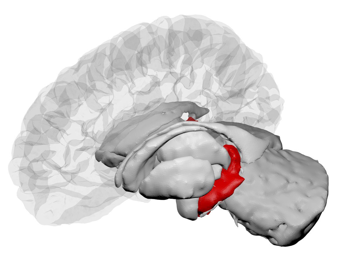
With the seizures growing worse through his twenties, Scoville offered him an experimental operation. He could not find a single point in the brain where the seizures began, so he removed a thumb-sized section of tissue from the inner part of each temporal lobe. In the paper he later wrote with the psychologist Brenda Milner, Scoville called it a frankly experimental operation.
The seizures that had ruled Molaison’s life largely stopped after the surgery, and his intelligence, personality and perception were unchanged. Within a short time, however, the doctors found that he forgot people almost as soon as they left the room. He could repeat a string of numbers straight away, but his recall of them fell away as the delay grew longer.
Read next
Why Apollo 11 Left Soviet Cosmonaut Medals on the Moon
What Brenda Milner Found at Hartford
Brenda Milner, a psychologist at McGill University in Montreal, made the first careful study of Molaison’s memory in 1955, two years after the operation. She and Scoville compared him with nine other patients who had undergone similar operations. Only those who had lost tissue from particular areas of the medial temporal lobe had trouble with memory, and the more tissue removed, the worse the loss.
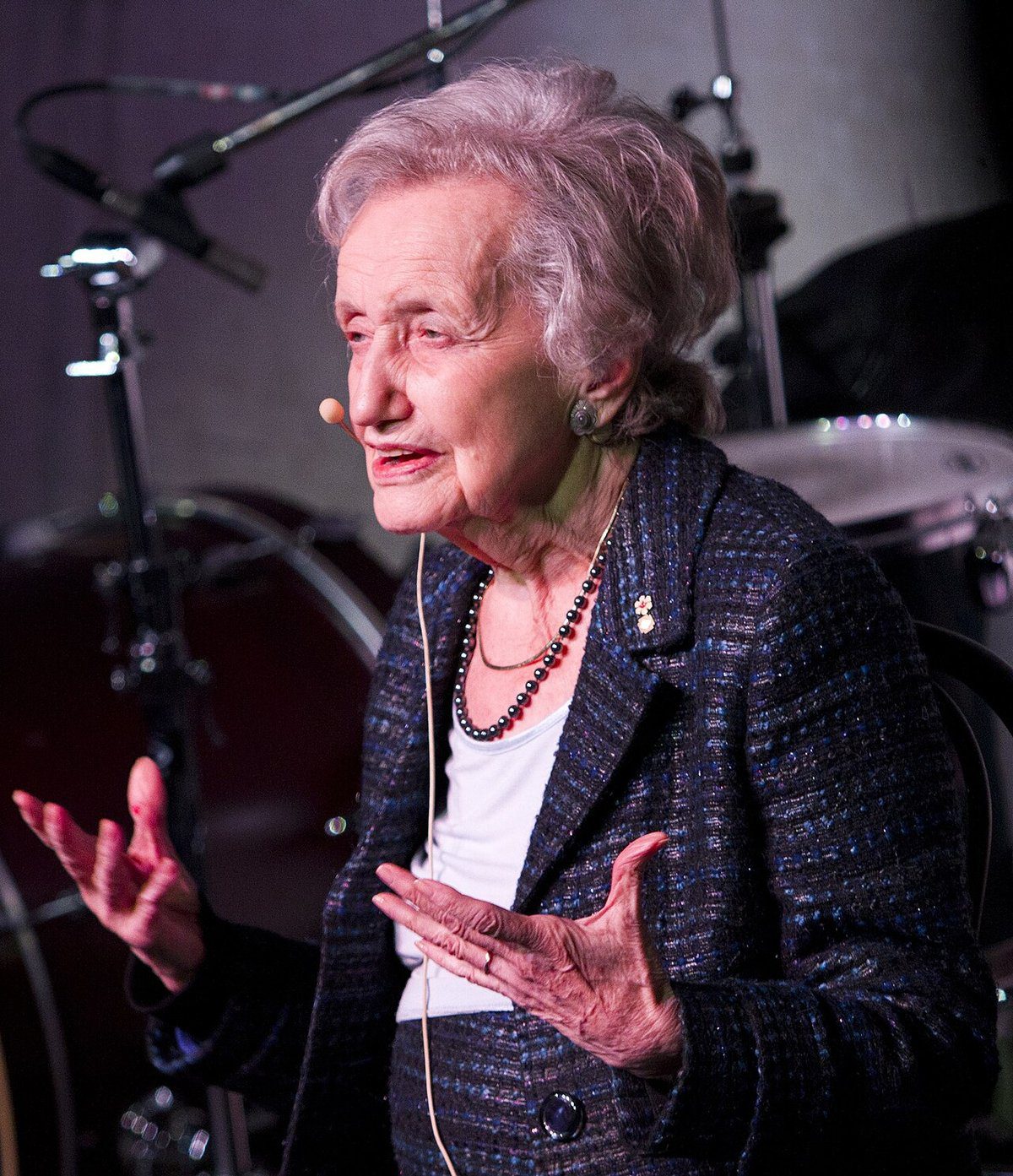
Their 1957 paper, titled “Loss of recent memory after bilateral hippocampal lesions,” concluded that the hippocampus was crucial for laying down memories of recent events. Milner recalled that every time she walked into the room to test him, it was as if they had never met.
Her most important test used a mirror and the outline of a star. Molaison was asked to trace the star while he could see his hand and the paper only in the mirror, which reverses every movement and makes the task hard for anyone at first. Over a series of sessions his tracing improved just as a healthy person’s would, even though he had no memory of having done the task before.
The mirror test showed that remembering facts and events depended on the tissue Scoville had taken, while learning a motor skill did not. Molaison could get better at something he did not know he had ever practiced, and the result became the evidence for two separate memory systems in the brain.
Decades With Suzanne Corkin
Suzanne Corkin, one of Milner’s graduate students at McGill, met Molaison in 1962, and two years later she moved to the Massachusetts Institute of Technology. She went on testing him there until his death, a span of about 46 years. He lived with his parents and other relatives for decades and moved into a nursing home in 1980, where he spent the last 28 years of his life.
Some knowledge did reach him despite the damage to his memory. In 1966 he drew the floor plan of his house, which surprised Corkin, who believed other parts of his brain had slowly taken in that knowledge. He knew that he had epilepsy, that he had had an operation and that his memory was poor, and he recognized people who had been famous before 1953.
Corkin described him as altruistic and said he took part in the research because he wanted to help other people. In a 1992 radio interview she asked him what he would do the next day, and he answered, “Whatever is beneficial.” That same year he agreed to donate his brain to MIT and Massachusetts General Hospital for study.
The Brain That Was Cut Into 2,401 Slices
Molaison died of respiratory failure on December 2, 2008, at a nursing home in Windsor Locks, Connecticut, at the age of 82. His full name was made public only after his death, and his brain was scanned at Massachusetts General Hospital and then taken to the University of California, San Diego.
There a team led by the researcher Jacopo Annese froze the brain in gelatin and, in December 2009, cut it into 2,401 thin slices. The two-day procedure was streamed live on the internet and watched more than 3 million times. Each slice was photographed so that the team could build a three-dimensional map of the whole brain at the level of single cells.
The results of that work were published in Nature Communications in January 2014. Annese said the aim was to revisit the original operation by dissecting it virtually, slice by slice, more than 60 years after Scoville had made his cuts.
Among the slices, the team found a small scar in the left orbitofrontal cortex, near the front of the brain, that no one had known about while Molaison was alive. Annese concluded that it was very likely made by Scoville during the 1953 operation.
More from Terribly Interesting
Sources
- BrainFacts.org (Society for Neuroscience)
- Benedict Carey, “H. M., an Unforgettable Amnesiac, Dies at 82,” New York Times, December 5, 2008 (text checked in a copy at
- McGill University newsroom
- UC San Diego, “H.M.’s Brain Yields New Evidence”
- AP via NBC News
- MIT News
- Scoville and Milner 1957, PubMed record
- Canadian Association for Neuroscience, Brenda Milner profile
- Undark
- MIT Brain and Cognitive Sciences